
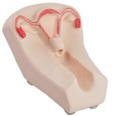
The DM-GP6624 demonstrates the anatomical structure of ovary, oviduct, uterus, cervix and vagina in coronal section. The transparent window cover lets trainees clearly observe insertion and placement of the intrauterine contraceptive device, and the placed IUD position can be checked after the procedure.
Key Features
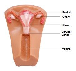
Extended Coronal Anatomy
Beyond the uterus, the model shows ovaries and oviducts, connecting IUD training with adnexal anatomy.
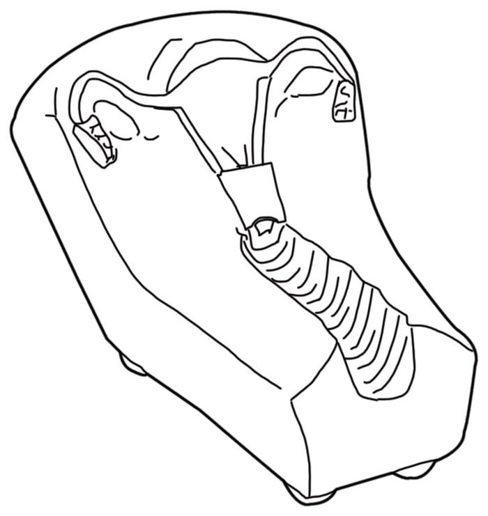
Clear Insertion View
The window cover exposes the whole insertion path from vagina through cervix into the cavity.
Placement Detection
After insertion, the IUD position inside the cavity is checked visually — correct fundal placement or misplacement.
Progressive Series
Model II bridges the basic coronal trainer (I) and the soft-tissue trainer (III) of the same series.
Standard Configuration
| NO. | Description | QTY |
|---|---|---|
| 1 | IUD Training Model II | 1 PC |
| 2 | User's Manual | 1 Copy |